
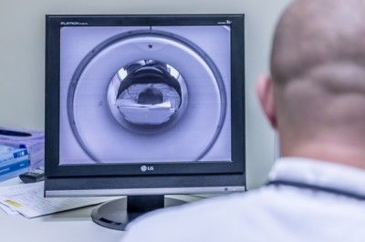
The prostate MRI
Once a patient has symptoms or complaints suggestive of prostate cancer, the patient is usually referred for an ultrasound. In most cases, this is also done if the patient has an elevated PSA level or the urologist finds evidence of prostate cancer during the annual screening exam.
Prostate MRI – underestimated alternative to “systematic” punch biopsy
If the ultrasound also provides indications for the suspicion of a prostate carcinoma, a biopsy is suggested by most practicing physicians. However, this procedure can lead to complications due to the non-sterile approach.
In addition, the hit rate of a conventional punch biopsy is remarkably low. Yet this could be spared over 50% of patients thanks to prostate MRI.
The velauf of a punch biopsy
In this procedure, the physician removes ten to twelve untargeted tissue samples with a needle. Needles are accessed through the rectum, which allows fecal matter to enter the bloodstream. Thus, the patient must always take a preventive antibiotic before this procedure.
Since prostate cancer is multifocal and the accessibility of the needles is limited, taking tissue samples is more of a shot in the dark. The low hit rate reflects these limitations.
Martin Löhr, head of the Clinic for Prostate Therapy in Heidelberg, says that the hit rate of the punch biopsy is at most 30 percent, which means that a patient often has to undergo several biopsies without getting certainty about his state of health.
A prostate MRI instead of biopsy
Compared to palpation and ultrasound, a multiparametric MRI examination enables early clarification of the cause of complaints and indications. While the other examinations can only detect changes, the MRI examination can distinguish between benign, malignant or inflammatory prostate diseases.
If it turns out that the patient only suffers from prostatitis or enlargement, he does not need any further clarifications. The patient is spared the biopsy (which is only necessary if cancer is suspected) and the attending physician can now initiate the correct treatment. The right decision of treatment requires thorough diagnostics.
If the MRI confirms the suspicion of prostate cancer, a biopsy is absolutely necessary. However, the above described 50 years old procedure of punch biopsy is not the only option. Because a prostate MRI also allows for a targeted biopsy.
Targeted biopsy thanks to live MRI
The MRI biopsy is very different from the punch biopsy. This diagnostic method offers maximum security and minimum consequences. In this case, the physician has live MRI images available while taking the tissue samples. This allows him to take targeted samples of the suspicious tissue.
The combination of targeted and systematically taken tissue samples leads to a phenomenal hit rate of 99 percent. The patient is thus offered maximum certainty. In addition, this biopsy is much more comfortable for the patient. Access is not via the rectum, but above the buttock region.
Thus, the patient does not need a preventive antibiotic or anesthesia, as this procedure is quick and painless. The patient is also spared an inpatient stay.
Conclusion
The MRI examination is a revolutionary milestone in medical diagnostics, allowing the structures and functions of tissues and organs to be examined in detail. With regard to suspected cancer, however, many practicing physicians still prescribe a punch biopsy, which is inaccurate and can also be very uncomfortable for the patient.
This procedure often has to be repeated several times, and in the end it may turn out that there is no prostate cancer. A detailed MRI examination can clarify at an early stage whether the disease is malignant.
If this is the case, you can still take advantage of an MRI and save yourself a punch biopsy. Because the biopsy with the help of live MRI is not only 99% correct, but also minimally invasive.